Resources

Showing 1-10 of 27
  • Media Image
    Paolo Longo, Ph.D., Gatan, Inc. Sample courtesy of Professor Jianfang’s group, Chinese University, Hong Kong Microscope courtesy of IBM, Fishkill, NY

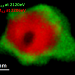
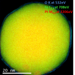
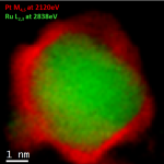
    Pd/Au alloys have attracted a lot of interest due to their resistance at high temperatures, and this explains their use in several fields, such as CO and hydrocarbon oxidation, synthesis of vinyl acetate monomer, hydrocarbon hydrogenation, and many others.

  • Media Image
    Paolo Longo, Ph.D., Gatan, Inc.

    Legend

    Cu L at 931 eV (grey); O K at 532 eV (red); N K at 401 eV (light blue); Ti L at 456 eV (green); Co L at 779 eV (blue)

  • Media Image
    Paolo Longo, Ph.D., Gatan, Inc. Sample courtesy of Dr. Jaco Oliver at NMMU, Port Elizabeth, South Africa Microscope courtesy of NMMU, Port Elizabeth, South Africa

    Analysis of the changes in local carbon structure caused by self-implantation of carbon into diamond and chemistry.

  • Media Image
    Paolo Longo, Ph.D., Gatan, Inc. Sample courtesy of Professor Gianluigi Botton at CCEM, Hamilton, ON, Canada Microscope courtesy of CCEM, Hamilton, ON, Canada Acknowledgement to Dr. Andreas Korinek at CCEM for helping set up microscope for experiment.

    EELS data taken using a probe and image-corrected FEI Titan TEM/STEM microscope equipped with X-FEG emission gun and a fully loaded GIF Quantum® ERS system. Au M4,5-edges at 2206 eV (red) and Pt M4,5-edges at 2206 eV (red).
    Methods

  • Media Image
    Paolo Longo, Ph.D., Gatan, Inc. Sample courtesy of Professor Gianluigi Botton at CCEM, Hamilton, ON, Canada Microscope courtesy of CCEM, Hamilton, ON, Canada Acknowledgement to Dr. M. Bugnet at CCEM for helping set up microscope for experiment.

    Pt/Fe nanocatalysts are among the most promising candidates for accelerating oxygen reduction reaction occurring at the cathode in a proton exchange membrane fuel cell.

  • Media Image
    Paolo Longo, Ph.D., Gatan Inc. Sample courtesy of Dr. Joshua Sugar at Sandia National Lab, Livermore, CA

    Methods

    FEI F20 TEM/STEM microscope; S-FEG emission gun; GIF Quantum® ER system; voltage: 200 kV; EFTEM SI mode from 680 – 730 eV; slit width: 2 eV ; step size: 1 eV; total exposure time:

  • Media Image
    Paolo Longo, Ph.D., Gatan, Inc. Sample courtesy of Dr. Giuseppe Nicotra at IMM-CNR, Catania, Italy Microscope courtesy of IMM-CNR, Catania, Italy

    Methods

    Probe-corrected ARM 200F TEM/STEM microscope; C-FEG emission gun; GIF Quantum® ER system; voltage: 60 kV; STEM mode; EELS low-loss spectrum (0 – 500 eV) exposure time: 0.01 ms; EELS core-loss spectrum (70 – 570 eV) exposure time: 10 ms; total exposure time:

  • Media Image
    Paolo Longo, Ph.D., Gatan, Inc. Sample courtesy of Professor Gianluigi Botton at CCEM, Hamilton, ON, Canada Microscope courtesy of CCEM, Hamilton ON, Canada Acknowledgement to Dr. Andreas Korinek at CCEM for helping set up microscope for experiment.

    Methods

  • Media Image
    Paolo Longo, Ph.D., Gatan, Inc. Sample courtesy of University of Glasgow Microscope courtesy of Dr. Yan Xin at Florida State University, Tallahassee, FL Acknowledgement to Dr. Toshiro Aoki at Jeol USA (now at ASU) for helping set up microscope for experiment.

    Dumbbell structure of GaAs on left hand side of color map is clearly resolved, and Ga and As atomic columns are visible. Interface region shows presence of Ga2O monolayer responsible for keeping Fermi level unpinned, which allows the electronic device to be turned on or off.

  • Media Image
    Paolo Longo, Ph.D., Gatan, Inc. Sample courtesy of University of Glasgow Microscopecourtesy of Professor Gerald Kothleitner, TU-Graz, Austria

    Methods

Pages