Media Image
Paolo Longo, Ph.D., Gatan, Inc.
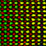
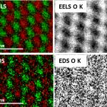
Sample courtesy of Dr. P. Rice and Dr. T. Topuria at IBM (Almaden), San Jose, CA
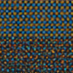
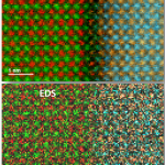
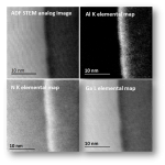
Microscope courtesy of Dr. Giuseppe Nicotra, IMM-CNR, Catania, Italy
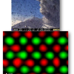
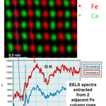
The lab where EELS maps were taken is situated at the slopes of Mount Etna in Sicily, Italy. Mount Etna is the highest active volcano in Europe. The EELS elemental maps were taken at high speed during major volcano eruption, as shown in the volcano webcam photograph above the maps.